Kasner eons in Lovelock black holes
Bueno P.; Cano P.A.; Hennigar R.A.; Li M.-D.
Physical Review D, Vol. 110, Num. 124015 (2024)
Article

El alumnado de 2º de primaria de la escuela La Pau de Sant Sadurní d’Anoia compartió en el Hub Social Barcelona los resultados de la investigación sobre el efecto del agua en las plantas, realizada a lo largo del curso de la mano de investigadores del ICTA-UAB.
Un equipo liderado desde el Centro de Biología Molecular Severo Ochoa del Consejo Superior de Investigaciones Científicas (CBM-CSIC-UAM) ha desarrollado organoides de oviducto bovino, pequeñas estructuras tridimensionales originadas en el laboratorio a partir de células de vaca extraídas del oviducto, el conducto que conecta el ovario con el útero. Estos organoides reproducen el funcionamiento del oviducto real, donde tiene lugar la fecundación y el desarrollo inicial del embrión en los mamíferos. Además, responden a hormonas como el estradiol y la progesterona, recreando los cambios hormonales del ciclo reproductivo. El hallazgo podría tener implicaciones para las técnicas de reproducción asistida. El trabajo se ha publicado en la revista Cellular and Molecular Life Sciences.
El trabajo liderado por el Dr. Vicente Pérez García del CBM-CSIC-UAM y el Centro de Investigación Príncipe Felipe (CIPF), en colaboración con la Universidad de Murcia, demuestra que estos organoides responden a hormonas reproductivas y generan secreciones biológicamente activas que reproducen las del oviducto in vivo.
El oviducto crea un entorno que cambia a lo largo del ciclo reproductivo y que es esencial para que el óvulo y el espermatozoide estén en las mejores condiciones para la fecundación y para que el embrión inicie su desarrollo. Este entorno está regulado por hormonas como el estradiol y la progesterona, que controlan la secreción de las moléculas necesarias para estos procesos.
Sin embargo, reproducir este sistema en el laboratorio ha sido un desafío importante. Los modelos tradicionales, como los cultivos en dos dimensiones o los esferoides, no logran imitar completamente la complejidad del oviducto real, lo que limita el estudio de la reproducción y el desarrollo de nuevas estrategias en reproducción asistida.
Frente a esto, los organoides desarrollados en este trabajo ofrecen una solución más precisa, ya que reproducen tanto la estructura como las funciones del oviducto. Los investigadores desarrollaron organoides a partir de células epiteliales que recubren el interior del oviducto bovino, encargadas de producir buena parte de las secreciones necesarias para la fecundación y el desarrollo temprano del embrión. Estos “organoides epiteliales” son capaces de organizarse en estructuras tridimensionales funcionales. Además, conservan la capacidad de responder a hormonas y de producir secreciones con un perfil parecido al del entorno natural.
Los resultados muestran que la exposición de estos organoides 3D al estradiol o la progesterona, dos hormonas que gobiernan el ciclo reproductivo, inducen cambios específicos en la expresión génica y en la composición de las secreciones que producen, representando fielmente las fases del ciclo reproductor.
“Estos organoides no solo mantienen la estructura polarizada del epitelio del oviducto, sino que también conservan su capacidad de responder a señales hormonales, lo que les permite generar un entorno muy similar al fisiológico”, explica Sergio Navarro-Serna, primer autor del estudio.
El término estructura polarizada se refiere a que las células se organizan de forma ordenada, según una orientación definida por sus funciones. En un epitelio secretor, las células suelen tener la zona por donde liberan secreciones (polo apical) orientada hacia el exterior o hacia una cavidad abierta, en este caso el lumen del oviducto. En los organoides, las células orientan su polo apical hacia el interior, es decir, hacia el hueco central, que constituye el lumen de la estructura. A diferencia del oviducto in vivo, donde el lumen está abierto hacia el ovario y el útero, los organoides presentan un lumen cerrado. Esta disposición supone una ventaja experimental, ya que permite que las secreciones se acumulen en ese espacio interno, facilitando su recogida y análisis de forma más eficiente. Gracias a esta organización polarizada, el tejido artificial deja de ser un simple conjunto de células y adquiere propiedades funcionales propias de un miniórgano de oviducto.
Uno de los hallazgos más relevantes del trabajo es que las secreciones generadas por estos organoides reproducen en gran medida la composición del fluido natural del oviducto. Además, los experimentos funcionales demostraron que estas secreciones, especialmente las generadas bajo estimulación con estradiol, son capaces de mejorar procesos fundamentales para los espermatozoides, como la capacitación y la reacción acrosómica, pasos esenciales para que puedan fecundar el óvulo.
La capacitación permite a los espermatozoides prepararse para la fecundación, cuando se exponen al ambiente especial del oviducto, enriquecido en proteínas y moléculas que les permiten moverse más rápido y de forma más eficiente, además de preparar su membrana para interactuar con el óvulo. Mediante la reacción acrosómica los espermatozoides pueden “romper” la capa externa que protege al óvulo (zona pelúcida) para entrar y fecundarlo, gracias a la liberación de enzimas desde una estructura situada en su cabeza llamada acrosoma. Ambos procesos son esenciales para la fecundación.
Así, este trabajo demuestra que las secreciones derivadas de los organoides no solo se parecen a las del oviducto en su composición, sino que también reproducen parte de su función biológica.
Estos resultados posicionan a los organoides del oviducto como una plataforma innovadora para estudiar la comunicación entre las células reproductoras y el entorno materno. Además, este modelo podría contribuir al desarrollo de nuevas estrategias para mejorar las técnicas de reproducción asistida, porque permitiría incorporar factores fisiológicos que actualmente no están presentes en los sistemas de cultivo convencionales.
“Este sistema ofrece una alternativa reproducible y controlada para estudiar procesos fundamentales de la reproducción, con potencial para mejorar la eficiencia y la calidad de los embriones generados in vitro”, concluye Vicente Pérez García.
Navarro-Serna, S., Romero-Aguirregomezcorta J., Hernández-Díaz N., Ferrero-Micó A., Coy P., Pérez-García V. (2026). Hormonally responsive bovine oviductal organoids recapitulate native oviductal secretions and enhance sperm capacitation. Cell Mol Life Sci. https://doi.org/10.1007/s00018-026-06200-6
La entrada Un modelo experimental en 3D recrea el entorno de la fecundación que podría mejorar las técnicas de reproducción asistida se publicó primero en Centro de Biología Molecular Severo Ochoa.

.jpg?itok=VXKT3y3c "jornada somma igualdad genero")
Faseeha Ayaz completed her undergraduate degree in Pharmacology at King’s College London before pursuing a DPhil in Cardiovascular Science at the University of Oxford. Her research focused on immunometabolism in diabetes and vascular function. In 2023, she joined Nature Communications, where she primarily handles papers on cardiovascular biology and disease, including preclinical, clinical, epidemiology, and machine learning manuscripts. She is also an advisory editor for npj Cardiovascular Health and a freelancing editor for Nature Cardiovascular Research and Communications Biology.
That’s a nice question to start with. I did my PhD at the University of Oxford in Cardiovascular Science. It was a wet-lab-based project in immunometabolism in atherosclerosis, so I was in the lab all the time. Unfortunately, I was doing my PhD during the COVID-19 pandemic, which made things particularly challenging as much of the scientific and research infrastructure had moved online. I found it difficult to be in the lab and to make the same connections.
More generally, I started my PhD loving lab work, but I ended it not enjoying it as much. I loved the lab environment, academia, and the science itself, but the manual aspects of lab work—and the fact that things don’t work out most of the time, which is just the nature of research—became tiring. So I moved into publishing, which is where I am now.
My current role is as close as you can be to science and academia without actually being in academia. It’s a very scientific environment as everyone has a PhD, and everyone is focused on publishing the best science and moving the field forward. The difference is that you’re not in a university setting and you’re not doing lab work.
Absolutely. One of the reasons I was attracted to this position is that my research project felt very niche. Academia often pushes you to become an expert in a very specific area, going deeper and deeper into it.
As an editor, however, you zoom out. You gain breadth of knowledge. I’m no longer focused only on immunometabolism in atherosclerosis. My job is to stay on top of many different fields—stroke, hypertension, diabetes, and more. You can’t make publishing decisions unless you understand what’s happening across the research landscape.
With this birds eye view, you start to see patterns; for example, metabolic reprogramming appearing in one area and then spreading into others. It’s fascinating to see how entire fields evolve.
Nature Communications is one of the largest journals in the world. As editors, we look for high-quality science and a clear conceptual advance. For example, if someone studies a particular protein in hypertension, we ask: has this been studied before in hypertension or in other cardiovascular diseases? What new knowledge does this bring? What is the impact on disease pathophysiology? Is it targetable? Is there human relevance? What models have been used?
We are looking for solid science. That may sound straightforward, but it represents years of work. These papers build the foundation of the field. Progress happens incrementally, and it is the collective effort of many researchers that ultimately advances science.
We usually aim for around three reviewers, with a minimum of two. With the previous example I mentioned, we would seek a reviewer with expertise in that protein, another with expertise in hypertension and the relevant models, and possibly a technical reviewer if specialized methods such as multi-omics are used.
Ultimately, the decision is made by the editor. We consider all reviewer comments, but it’s not about counting votes. It’s about evaluating the strength of the arguments and how they affect the quality of the manuscript.
There are several major unanswered questions. One example is heart failure with preserved ejection fraction, which is a highly heterogeneous disease, and key questions remain: what are the patient subgroups? What are the phenotypes? What is the metabolic component? It’s increasingly viewed as a metabolic disease, but the molecular mechanisms are still unclear. Papers addressing these questions are particularly exciting.
Another area is GLP-1 agonists. They have revolutionized obesity treatment, but their cardiovascular effects are still not fully understood. Clinical measures often focus on weight loss or cholesterol levels, but not necessarily on outcomes like all-cause mortality.
Understanding how these therapies work mechanistically and whether they truly impact long-term outcomes how is something I find particularly interesting.
Yes, one comes to mind. The authors conducted a mechanistic clinical trial in which they studied “the athlete’s paradox”, which refers to both athletes and diabetic patients having increased intramyocellular fat levels despite opposing insulin sensitivity and cardiovascular disease risk. After placing the diabetic patients on endurance training and the athletes on deconditioning, they found that diabetic patients had higher unsaturated intramyocellular fat, lower palmitate kinetics. This was reversed with following endurance training.

It was a fascinating question and gives you that sense of, “this is really interesting”—it has direct human relevance and is a very clean, well-designed study. It addresses a question that researchers have been asking for years, and it’s a fascinating question in itself.
For me, an elegant paper is one that answers a complex and important question in a very simple way. When you see it, you think, “of course—that makes perfect sense.” But it only seems obvious in hindsight.
I think article processing charges at Nature Communications, as well as across the Nature portfolio, can be considered quite high. Many Nature journals offer hybrid model where APCs are only charged if the article is open access. At Nature Communications, all our content is open access.
What the company tries to do is offer fee waivers. If you are from a less economically developed country—and there is a defined list—you may receive a 50%, 70%, or even a full waiver of the APCs (Article Processing Charges). Spain, for example, is not included.
We also always encourage researchers to submit regardless of APCs, because editors are not involved in costs at all. I personally have nothing to do with that process—once a paper is accepted, it moves into production.
We are also advised to reassure authors not to be discouraged by APCs and to submit their work. In some cases, solutions may be found after acceptance. Overall I would say the editors are very much on the researchers’ side. The financial aspects are handled at the company level.
I think there may be a shift away from the US especially as other regions, including China, India and parts of the Middle East, are investing heavily in research. Funding is essential for science. I personally see many submissions from China, as well as from the US and Europe. China’s investment in research is very strong, and both the volume and quality of research are increasing significantly.
AI is becoming integrated into science. It is already being used to detect fraudulent papers and paper mills. AI is not used for editorial decisions, which remain entirely human. Researchers and reviewers can use AI for writing support, but not to generate data, and its use must be declared. AI may democratize writing, especially for non-native English speakers, but it may also narrow the scope of research. It is clearly reshaping how science is conducted.
Editors don’t directly use AI to detect AI-generated content. However, this may be done as part of checks before or after the editor sees the paper. If there are any red flags raised, we liaise with other teams in the company who can assess this for us.
Every journal also has its own focus and audience so understanding what each journal is looking for is key. Reading the journal and speaking with editors helps with this. For example, for pre-clinical work, at Nature Communications we look for strong mechanistic insight and depth. This kind of mechanistic focus may differ from journals such as Circulation or the European Heart Journal.
Editorial decision making is never a tick-box exercise and involves many different parameters. That being said, there are useful guidelines which I will share during my talk so that, when researchers are preparing their papers, they can think about whether additional elements or further validation might strengthen their work.
The simplest way to understand if the journal is the right home for your paper is to submit it and receive feedback. We also offer transfer options within the Nature portfolio, helping authors find a suitable journal. Otherwise, reviewing the journal’s content and consulting editors can help.

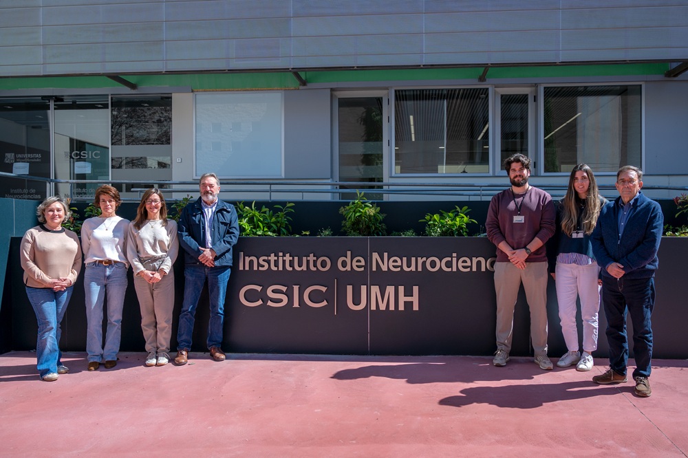
Photo: Researchers from the Institute for Neurosciences: Francisca Almagro, Raquel García López, Ana Pombero, Daniel Garrigós, Marta Martínez, and Emilio Geijo. Source: IN UMH-CSIC
YouTube video: https://youtu.be/sSX7WGTHlew?si=GEIXBYK-S2q3OHsC
Amyotrophic lateral sclerosis (ALS) is a neurodegenerative disease that causes the progressive loss of motor neurons, which in most cases leads to respiratory failure within three to five years after diagnosis. In this context, a team from the Institute for Neurosciences (IN), a joint centre of the Miguel Hernández University (UMH) and the Spanish National Research Council (CSIC), has identified that a cellular ‘selective cleaning system’ for proteins: chaperone-mediated autophagy, is significantly reduced in patients, making it a potential therapeutic target to slow disease progression.
The study, published in Acta Neuropathologica Communications, involved the participation of the UMH Sports Research Centre and the Pascual Parrilla Murcia Institute for Biosanitary Research (IMIB). In this work, the team explored in greater depth the role of this cellular mechanism, which is responsible for the selective removal of damaged proteins. Its proper function is essential to maintain neuronal homeostasis, and its dysfunction may promote the accumulation of toxic proteins, one of the hallmark features of the disease.
“ALS is a devastating disease whose cause remains unknown in the vast majority of patients, which greatly hampers the development of effective treatments”, explains Professor Salvador Martínez, director of the Neurobiology of Mental, Neurodegenerative, and Neuro-oncological Diseases Laboratory at the IN UMH-CSIC. He adds that “identifying cellular mechanisms directly involved in neuron survival is a key step towards advancing new therapeutic strategies”.
An essential system for neuronal survival
Motor neurons are particularly vulnerable cells in ALS. In more than 90% of cases, these cells accumulate a protein called TDP-43 outside its normal location, forming toxic aggregates. The body has mechanisms to prevent this accumulation, including autophagy, a cellular ‘cleaning and recycling’ system. However, not all types of autophagy work in the same way. While macroautophagy acts as a general waste disposal system, chaperone-mediated autophagy is highly selective and is responsible for degrading specific proteins, such as TDP-43.
To analyze this process, the researchers used spinal cord tissue from patients enrolled in clinical trials conducted by the IN UMH-CSIC and IMIB teams, as well as control samples from donors without the disease. Using immunohistochemistry and immunofluorescence techniques, they assessed the presence of LAMP2A, a key protein that serves as an indicator of the activity of this type of autophagy.
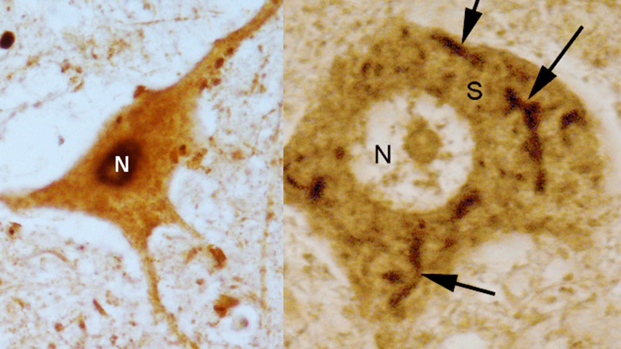
Spinal cord motor neurons. Left: healthy motor neuron, in which the TDP-43 protein (in black) is localized in the cell nucleus (N). Right: motor neuron from an ALS patient, in which TDP-43 appears abnormally accumulated in the cytoplasm of the cell body (S). Source: Acta Neuropathologica Communications.
The results show that healthy motor neurons display high activity of this system, whereas in patients with ALS, this activity is markedly reduced. “These findings indicate that chaperone-mediated autophagy activity is clearly decreased in motor neurons from ALS patients”, explains Daniel Garrigós García, first author of the paper. In this regard, Martínez highlights: “In our study, we have shown that motor neurons require very high levels of chaperone-mediated autophagy to survive. When this mechanism declines, as occurs in ALS, these are precisely the cells that are first affected and eventually die”.
Donations that drive research
This study also shows that the observed alterations are specific to cellular protein clearance and recycling processes and that they present significant differences between patients and controls. “We have been able to observe this mechanism directly in human tissue, something we had not achieved in animal models”, says Martínez, who highlights that the study was made possible thanks to the altruistic donation of tissue by patients and their families, which is essential for advancing ALS research.
Based on these findings, the researchers suggest that this mechanism could become a new therapeutic target. “Our goal is to try to modulate this pathway to increase its activity”, explains the professor, adding that this discovery opens the door to the development of strategies aimed at slowing disease progression, although they are still in the early stages of research.
This work has been made possible thanks to funding from the Spanish State Research Agency, through the “Severo Ochoa” Programme for Centres of Excellence in R&D; the Ministry of Science, Innovation and Universities; the Prometeo Programme of the Generalitat Valenciana; the Instituto de Salud Carlos III (Advanced Therapies Network – TERAV); and the Next Generation EU programme within the framework of the Recovery, Transformation and Resilience Plan. It also received key support from the UMH Chair Gregoria Ramos Gil on ALS.
Chaperone mediated autophagy is deficient in spinal motoneurons of ALS patients with TDP-43 proteinopathy. Garrigos, D., Martinez-Morga, M., Pombero, A., García-Lopez, R., Pastor, D., Riquelme, D., Blanquer, M., Iniesta, F., Valdor, R., Geijo-Barrientos, E., Hargus, G., Moraleda, J.M and Martínez, S. Acta Neuropathologica Communications, 2026; 14, 67.
Source: Institute for Neurosciences UMH-CSIC (in.comunicacion@umh.es)
La entrada A study from the Institute for Neurosciences UMH-CSIC identifies a key mechanism in the degeneration of motor neurons in ALS se publicó primero en Instituto de Neurociencias de Alicante.

The document announces the prioritised applicants.