- A Chinese-Spanish collaboration led by Lola Martin-Bermudo revealed the role of the nidogen protein in Drosophila
- The work details the effect of the loss of function of the domains of nidogen in different tissues and various stages of development
- The findings challenge previously held ideas in the field of development and morphogenesis
Every tissue of the human body has a lining called the basement membrane. This structure has a role in the separation of internal and external body surfaces and is essential for the proper . This membrane is composed of thin layers of cells having specialized external cell matrices, which are structures outside, but associated to these cells.
The matrices are in fact scaffolds of proteins arranged in a mesh-like fashion their main components being proteins called laminin and “type IV” collagen, linked together by other proteins known as proteoglycans and nidogen (Ndg). In the described matrix ensemble, the function of nidogen has remained a controversial aspect, even though it was yet known that it plays a key role during organogenesis and late embryo development (particularly, regarding heart or lung development).
A chance encounter in the Matrix
During a Matrix meeting in the past, an encounter took place between researchers Jose C. Pastor-Pareja (School of Life Sciences, Tsinghua University, Beijing, China) and Lola Martin-Bermudo (GEM-DMC2, CABD, Seville, Spain), by which they realised that they were both researching about the nidogen protein.

About that momentous encounter, Martin-Bermudo declared that ‘it was surprising to learn that Pastor-Pareja’s lab was also trying to isolate a nidogen mutant in Drosophila’. As they discussed on their research, the fact that in fruit fly there is only one nidogen variant while in vertebrates there are 2 isoforms of the protein also was highlighted, raising further questions for the future.
They found, the information from experiments in cell culture and animal models such as mouse and the worm Caenorhaditis elegans were clearly contradictory. On the one hand, the results from cell culture indicated that nidogen was essential to link the collagen and laminin matrix. Nidogen, additionally, conferred a higher stability to the matrix. Despite this, C. elegans and mouse mutants with impaired nidogen protein appeared nonetheless to be viable.
This apparent paradox led the researchers to wonder if cell culture experiments were reflecting adequately the situation occurring in the full organism. This could be pinpointing the requirement for a physical support in the use of cell cultures for obtaining solid scientific conclusions in this context.
A deal is struck… with positive outcomes
The mutual interest, but also heavy competition in their research field led Pastor-Pareja and Martin-Bermudo’s lab to join efforts to dissect the role of nidogen using the fruit fly Drosophila. A big effort was launched to map the parts of the nidogen protein interacting with collagen and laminin, as nidogen was thought to be a crucial component of the matrix.

Both labs isolated in parallel mutants with severely impaired, mutated nidogen protein. The mutant from Martin-Bermudo’s lab contained a defect in a crucial domain for the binging of nidogen to the other main proteins of the matrix. Detailed analysis of nidogen function during development and its role linking laminin and collagen took place. The mapping of the key domains involved in the binding showed that most of the conclusions previously drawn from cell culture experiments were not correct.
The work, published in PLOS Genetics, challenged several ideas previously held in the field of development and morphogenesis. One of its main conclusions was that several of the properties of nidogen are tissue-specific. It follows that in the hierarchy of the matrix assembly, the properties of that matrix are not only dependent on collagen and laminin but also on the nidogen protein. By the exhaustive analysis of nidogen function, it became clear that the function and the interaction of the components of the matrix depends on the tissue and moment of development.
Image credits:
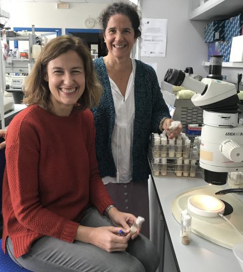
Picture of researchers kindly provided by GEM-DMC2, CABD.
Fly on yellow flower picture dowloaded from Wikipedia (Portuguese) and licensed via a Attribution-ShareAlike 4.0 International (CC BY-SA 4.0).
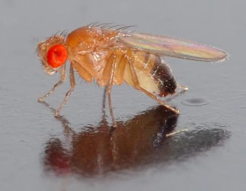
Drosophila melanogaster picture downloaded from Wikimedia Commons and licensed via an Attribution-ShareAlike 2.5 Generic (CC BY-SA 2.5).
